
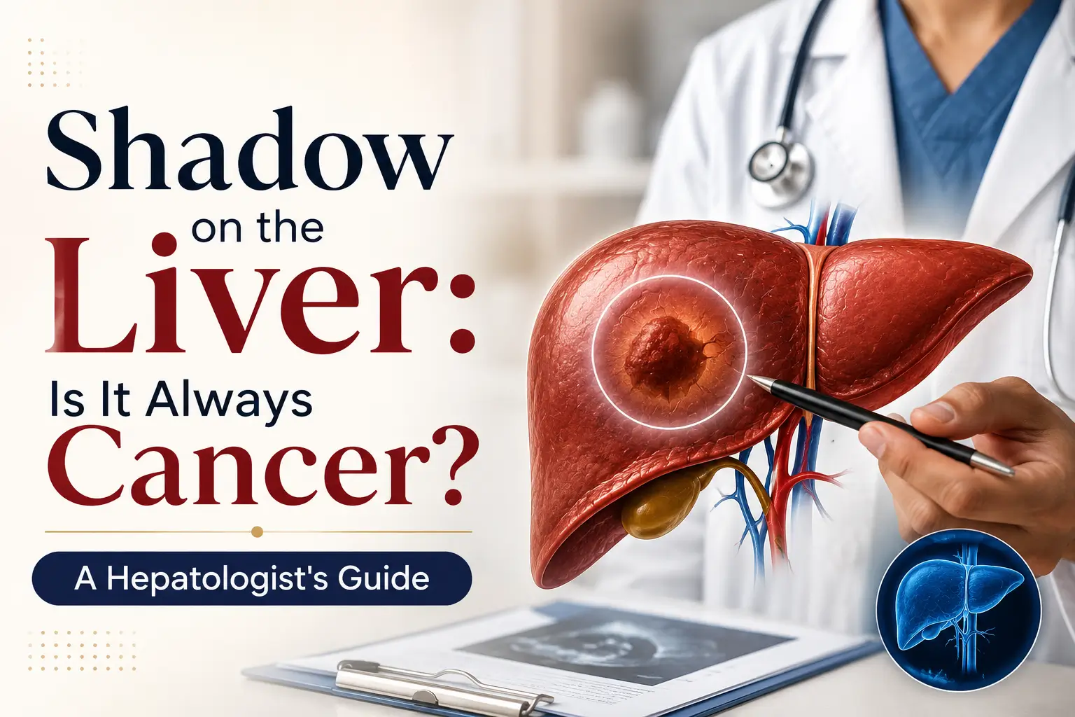
If you have been told there is a shadow, spot, or lesion on your liver perhaps found unexpectedly on an ultrasound or CT scan the first question that usually comes to mind is a frightening one: is this cancer?
The honest answer, in most cases, is no. The great majority of shadows and lesions found on liver scans turn out to be benign. Some are so common they need no treatment at all. Some are entirely harmless features that many people carry through life without ever knowing. Only a minority of liver lesions are cancerous.
That said, we take every liver finding seriously at our Harley Street clinic because although most are benign, the ones that aren’t need to be identified quickly. This guide walks you through what different liver lesions actually mean, when a finding is concerning, and how a hepatologist works out what’s going on.
No — and this is worth stating clearly. Studies suggest that when an incidental liver lesion is found on routine scanning, the great majority turn out to be benign. In patients without underlying liver disease or a history of cancer, it is even less likely a liver lesion is cancerous.
The medical term for these findings is ‘focal liver lesions’. They cover a wide range of things, from simple cysts (which are entirely harmless) through to primary liver cancer (which is serious and need prompt treatment). What matters is not the fact that something has been seen — it’s what that something turns out to be.
This is one of the situations where a consultant hepatologist review really matters. A specialist liver consultation combines your scan images, medical history, and further testing to give you a definitive answer — usually within days rather than weeks.
There are more types of liver lesion than most patients realise. Understanding the range helps put a new finding in perspective.
Type of Lesion | Cancerous? | What It Is |
Simple liver cyst | No | A fluid-filled sac. Very common, often present from birth. Rarely causes problems and usually needs no treatment or follow-up. |
Haemangioma | No | A cluster of small blood vessels. The most common benign liver lesion. Usually found by chance and left alone, unless large. |
Focal Nodular Hyperplasia (FNH) | No | An overgrowth of normal liver tissue. More common in women. It is benign. |
Hepatic adenoma | Usually no | A benign tumour, sometimes linked to hormonal contraceptives. Occasionally needs treatment or monitoring as it can transform to cancer. |
Regenerative nodules | Not directly | Small nodules that form in cirrhotic livers. Not cancer themselves but do require monitoring. |
Hepatocellular carcinoma (HCC) | Yes | The most common primary liver cancer. Usually occurs in a liver already affected by chronic disease (cirrhosis, hepatitis B/C, MASLD). |
Cholangiocarcinoma | Yes | Bile duct cancer. Less common but requires prompt specialist assessment and treatment. |
Secondary (metastatic) tumour | Yes | Cancer that has spread from elsewhere in the body (bowel, breast, lung). More common than primary liver cancer. |
The key point is that the majority of the lesions on this list are green — that is, benign. Even hepatic adenomas and regenerative nodules, which need monitoring, are not the same thing as cancer. A hepatologist’s job is to identify which category a specific finding belongs to, using a combination of imaging, blood tests, and clinical context.
When a lesion does turn out to be malignant, it falls into one of three categories. Each behaves differently and is managed differently.
HCC is the most common primary liver cancer — ‘primary’ meaning it starts in the liver itself. HCC almost never arises in a healthy liver. It occurs on a background of chronic liver disease, most often cirrhosis. The main risk factors in the UK are long-standing hepatitis B or C, heavy alcohol use over years, and advanced fatty liver disease (MASLD).
This background matters because it changes how we investigate findings. In a patient with cirrhosis, a new liver nodule is treated with high suspicion. In a patient without any known liver disease, the same finding is more likely to be benign.
This is cancer of the bile ducts — the small channels that carry bile from the liver to the intestine. It’s less common than HCC in the UK, but has been rising modestly over the past two decades. It sometimes presents with jaundice (yellowing of the skin and eyes) because the bile ducts become blocked.
Secondary cancer is where a tumour originating elsewhere in the body has spread to the liver. In practice, this is actually the most common form of liver cancer overall in the UK. Bowel cancer is pne of the most frequent sources, alongside breast, lung, and pancreatic cancers. Secondary liver cancer is managed by the specialist team treating the original cancer.
The most important thing to know about liver cancer is that in the early stages, most people have no symptoms at all. This is why surveillance is so important for patients at higher risk — we catch cancers early not because patients notice symptoms, but because we look for them proactively.
When symptoms do appear, they tend to be vague and easily attributed to other causes. That said, the following symptoms warrant seeing a doctor, particularly if they are new, persistent, or occurring together:
None of these symptoms individually mean cancer. Each has many possible explanations, most of them not serious. But they do mean it is worth seeing a doctor to work out what is going on.
Liver cancer is uncommon in the general population but far more common in specific groups. The vast majority of primary liver cancers occur in patients with one or more of the following:
For patients with cirrhosis, six-monthly liver ultrasound surveillance is recommended precisely because early detection makes a genuine difference to outcomes. If you have cirrhosis and you are not being invited for routine surveillance, that is worth raising with a hepatologist.
Similarly, patients with advanced fatty liver disease (MASLD) should have their fibrosis stage assessed, since it is fibrosis and cirrhosis — rather than fatty liver itself — that carries the cancer risk.
When a shadow or lesion is found on an initial scan, the investigation typically follows a clear pathway:
A hepatologist reviews your history: any known liver disease, hepatitis exposure, alcohol history, family history, medications, and existing scan reports. This context alone often narrows the range of possibilities considerably.
Standard liver blood tests plus tumour markers, most commonly alpha-fetoprotein (AFP). These are used alongside imaging — no blood test alone diagnoses or rules out liver cancer.
Multiphase CT or MRI with contrast provides detailed information about the appearance and behaviour of a lesion. In parallel, a FibroScan helps assess the underlying condition of the rest of the liver, which is critical context for interpreting any lesion.
In modern hepatology, biopsy is not routine. Advanced imaging can characterise most lesions definitively. Biopsy is reserved for cases where the imaging remains unclear, or where confirming the specific tissue type would change treatment.
When liver cancer is confirmed, staging determines the best treatment approach. The system most commonly used in the UK and Europe is BCLC (Barcelona Clinic Liver Cancer). It classifies cancer not just by the size and number of tumours, but also by liver function and overall health — because both matter enormously for treatment.
The important point about BCLC is that it emphasises that early-stage liver cancers are often curable. This is exactly why surveillance in high-risk patients matters.
Treatment is highly individualised and depends on the stage, the underlying liver condition, and each patient’s overall health. In broad terms, the main options include:
Removing the part of the liver containing the tumour. Suitable for smaller cancers in patients with well-preserved liver function. The liver has a remarkable capacity to regenerate, making this a curative option in many early cases.
For patients meeting specific criteria — typically early-stage cancer in the context of cirrhosis — transplant offers the best long-term outcomes. It treats both the cancer and the underlying liver disease at once.
Techniques such as radiofrequency ablation or microwave ablation destroy the tumour using heat, without removing tissue surgically. Effective for smaller tumours and often used in patients who aren’t ideal surgical candidates.
These treatments deliver chemotherapy or radiotherapy directly to the tumour through the blood vessels feeding it, sparing the rest of the liver. Used for intermediate-stage disease.
Immunotherapy and targeted drug therapies have transformed the outlook for advanced liver cancer over the past decade. New combinations are now standard first-line treatment and are showing meaningful survival improvements.
For patients at any stage, symptom management, nutritional support, and quality-of-life care are integral parts of treatment — not just something considered at the end.
If you are in one of the higher-risk groups — cirrhosis, hepatitis B or C, advanced fatty liver disease with fibrosis, or hereditary haemochromatosis — you should have routine liver cancer surveillance. Current UK and international guidelines recommend:
This is genuinely one of the most valuable things we do in liver medicine. Cancer detected at surveillance is almost always caught at a stage where curative treatment is possible. Cancer detected because of symptoms tends to be caught later, when curative options are more limited. The difference in outcomes between these two situations is substantial.
No. In most cases, an incidentally found liver lesion is benign. Simple cysts, haemangiomas, and focal nodular hyperplasia together account for most findings.
Yes, at early stages. When liver cancer is caught at early stages typically through surveillance in high-risk patients — curative treatment is often possible through resection, transplant, or ablation. Later-stage cancers may not be curable but can often be controlled effectively with modern treatments.
Not always. In many cases, advanced imaging (multiphase CT or MRI) is sufficient to diagnose liver cancer without a biopsy. Biopsy is reserved for cases where the imaging is uncertain or where confirming the specific tissue type would change management.
This varies significantly between individuals and tumour types. Some hepatocellular carcinomas double in size in a few months; others grow much more slowly. This variability is one of the reasons regular surveillance (rather than one-off assessment) matters for high-risk patients.
Liver cancer itself is not typically hereditary, but some risk factors are — for example, hereditary haemochromatosis (iron overload) can increase risk over decades. If a first-degree relative has had liver cancer, it is worth discussing your own liver health with a hepatologist, particularly if you have any additional risk factors.
Fatty liver disease itself doesn’t turn into cancer, but it can progress to fibrosis and cirrhosis over years, and cirrhosis carries a real cancer risk. This is why detecting and reversing fatty liver disease early matters. Our guide to fatty liver disease covers this in detail.
In everyday language, patients often say ‘shadow’ when a radiologist has described a ‘lesion’, ‘nodule’, or ‘mass’. Medically, these terms mean the same thing: an area of the liver that looks different from the surrounding tissue on imaging. The word doesn’t tell you whether it is benign or malignant — that determination requires further assessment.
The most important message to take from this article is that a shadow, lesion, or mass on the liver is not the same thing as cancer. In most cases, it isn’t cancer at all. But it is worth having a proper answer, and that usually means combining your scan images with expert clinical review, further imaging where needed, and a hepatologist’s judgement.
At Leaders in Liver Health, our consultants provide rapid, expert assessment of liver lesions — usually with a clear diagnosis within days. You can book a private liver specialist consultation at our Harley Street clinic. Same-week appointments are usually available. Call us on 020 4621 0765 to discuss your case.
hepatocellular carcinoma. Journal of Hepatology, 2018.
National Institute for Health and Care Excellence (NICE). Liver disease: assessment and management guidance.
Reig M et al. BCLC strategy for prognosis prediction and treatment recommendation: The 2022 update. Journal of Hepatology. 2022;76(3):681-693.
British Society of Gastroenterology (BSG). Guidelines on the management of hepatocellular carcinoma.
British Liver Trust. Primary liver cancer and secondary liver cancer — patient information.
Cancer Research UK. Liver cancer statistics — UK incidence, survival, and risk factors.
This article was reviewed by Apostolos Koffas, Consultant Hepatologist at Leaders in Liver Health, (GMC: 7360520). [Brief 2-line bio mentioning hospital affiliations, areas of expertise, and years of experience]. Read more on our team page.
Prevention is the best medicine, but knowing where to start can be challenging. Our liver specialists provide evidence-based prevention advice tailored to your individual circumstances.
Take control of your liver health with expert guidance.
WhatsApp us
Fill out the form below to schedule a consultation with our liver health specialists. Our team will review your information and contact you within 24 hours to arrange an in-person or virtual consultation. This helps us understand your concerns and offer the best personalized care for your liver health.